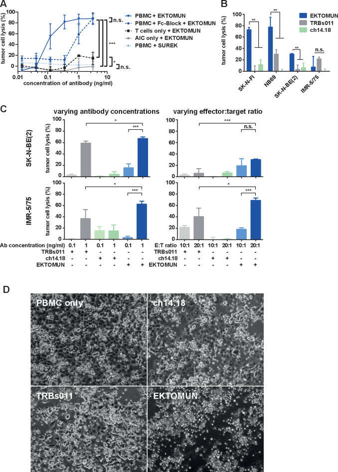
Figure 2.
EKTOMUN mediates a cytotoxic effect against neuroblastoma cell lines in the presence of human PBMCs. (A) The NB69 neuroblastoma cell line was stably transduced with a GFP_ffluc construct and cocultivated with T cells, effector:target (E:T)=7:1; accessory immune cells (AIC), E:T=3:1; or PBMCs that include both T cells+AICs, E:T=10 (7+3):1, from the same healthy donor. Cocultures were treated with the trAbs EKTOMUN or SUREK in rising concentrations with or without FcγR-block. Tumor cell lysis was determined by bioluminescent flux relative to an untreated coculture after 72 hours. (B) The MYCN-non-amplified neuroblastoma cell lines SK-N-FI (low GD2 expression) and NB69 (medium GD2 expression) and the MYCN-amplified cells lines SK-N-BE(2) and IMR-5/75 (both high GD2 expression) were cocultured as described in (A) and treated with the trAbs EKTOMUN or TRBs011 or with the monoclonal ch14.18 antibody. Tumor cell lysis was determined by bioluminescent flux relative to an untreated coculture after 72 hours. (C) SK-N-BE(2) and IMR-5/75 cells were cocultured as described in (A) and treated with the trAbs EKTOMUN or TRBs011 or with ch14.18 at indicated concentrations at an E:T ratio of 1:10 (left panels) and at indicated E:T ratios at an antibody concentration of 0.1 ng/mL (right panels). Tumor cell lysis was determined by bioluminescent flux relative to an untreated coculture after 72 hours. (D) NB69 cells were cocultured as described in (A) and treated with either the trAbs EKTOMUN, TRBs011, ch14.18 at 0.1 ng/mL, or without antibody. Micrographs were taken after 72 hours. Results are pooled medians of technical triplicates of three independent experiments. Student’s t-test; *p<0.05; **p<0.01.

