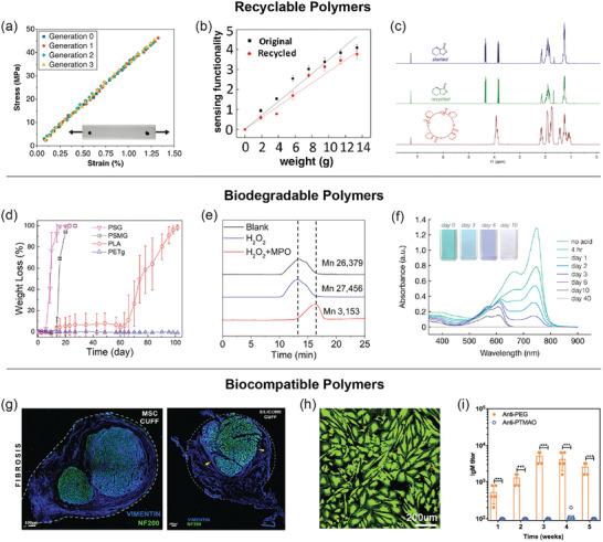
Figure 2.

Typical characterization methods used for recyclable, biodegradable, and biocompatible polymers. a) Indistinguishable stress–strain curves of cyclic poly(phthalaldehyde) (cPPA) under quasi‐static tensile loading over three generations of recycling. Adapted with permission.[ 22 ] Copyright 2019, American Chemical Society. b) Similar sensing performance of the polyimine‐based tactile sensor before and after recycling. Adapted with permission.[ 23 ] Copyright 2018, AAAS. c) Overlays of 1H NMR spectra of starting (blue) and recycled (green) lactone‐based monomers and polymer (red), which display identical chemical shifts (ppm) for the starting and recycled monomers. Adapted with permission.[ 25 ] Copyright 2018, AAAS. d) Weight loss profiles of polyesters PSG and PSG show significant degradation compared to controls PLA and PETg at 50 °C in phosphate‐buffed saline (pH = 7.4). Adapted with permission.[ 38 ] Copyright 2020, American Chemical Society. e) GPC traces of the original and degraded SPNs show a reduction of molecular weight based on retention time after treatment with both H2O2 and myeloperoxidase. Adapted with permission.[ 20 ] Copyright 2017, Springer Nature. f) UV–vis absorption spectra of a semiconducting imine‐based polymer solution under acidic conditions decreased over 40 d, demonstrating a loss of conjugation. Adapted with permission.[ 4 ] Copyright 2019, American Chemical Society. g) DIC microscopy images of nerve cross sections with shape memory polymer‐based MSC showed normal nerve fibers (green), while that with a silicone cuff showed nerve compression by fibrotic tissue ingrowth (arrowheads). Dotted lines indicate the relative positions of the MSC and silicone devices. Adapted with permission.[ 49 ] Copyright 2018, Springer Nature. h) LIVE/DEAD images of cells cultured on PAA‐rGO hydrogel show flourishing cell growth (green) and the absence of cell death (red). Adapted with permission.[ 51 ] Copyright 2018, Elsevier. i) The titer of PEG‐ and PTMAO‐specific immunoglobin M (IgM) in mice sera was detected with ELISA tests, demonstrating minimal immunogenicity of PTMAO. Adapted with permission.[ 52 ] Copyright 2019, AAAS.
