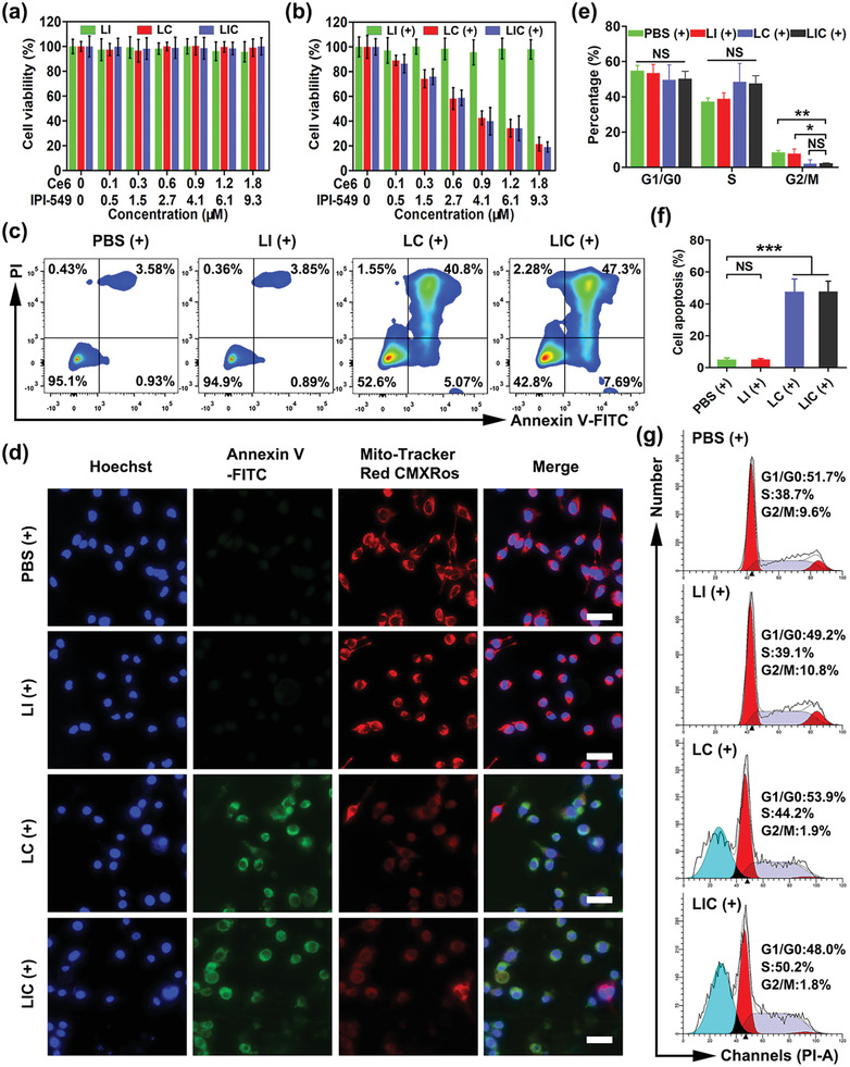
Figure 2.

In vitro killing effects of nanodrugs on CT26 cells. In vitro cytotoxicity of LI, LC, and LIC a) without or b) with light irradiation (660 nm, 0.8 W cm−2, 1 min) (n = 4). c) CT26 cell apoptosis determined by flow cytometry. d) Fluorescence images of Annexin‐V staining (green) and staining for change in mitochondrial membrane potential (red). Scale bar: 25 µm. e) Statistical analysis of cell cycle distribution and f) apoptotic rate of CT26 cells in different groups (n = 3). g) Representative plots of CT26 cell cycle distribution. (+) represents 660 nm (0.8 W cm−2, 1 min) light irradiation. *p < 0.05, **p < 0.01, ***p < 0.001, NS: no significant difference.
