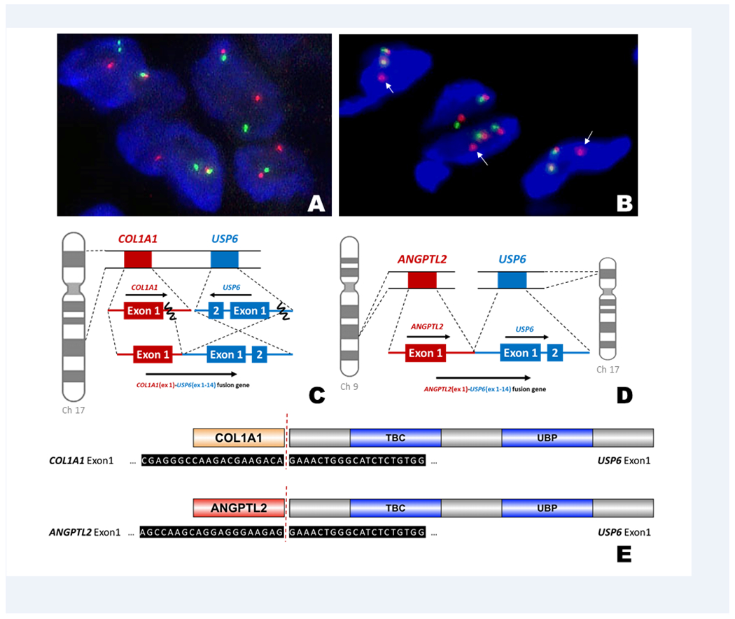
FIGURE 1.

(A) Fluorescence in situ hybridization (FISH) analysis with break-apart probes flanking the USP6 gene (5’ – green, 3’ – orange) showed a split of the green and orange signals, indicating disruption of USP6. (B) Two normal signals and a single 3’ (arrow) signal indicative of USP6 gene rearrangement was seen in case 3. (C) This schematic diagram represents the most common fusion pattern in our series of MO-like ABCs (6/7 cases), with breakpoints within exon 1 of COL1A1 (17q21-1722) and exon 1 of USP6 (17p13). (D) A novel fusion between ANGPTL2 (9q33) and USP6 was found in case 3. (E) The COL1A1-USP6 and ANGPTL2-USP6 fusion products included the promoter region of the COL1A1 and ANGPTL2 and the entire coding sequence of USP6.
