
FIGURE 4.
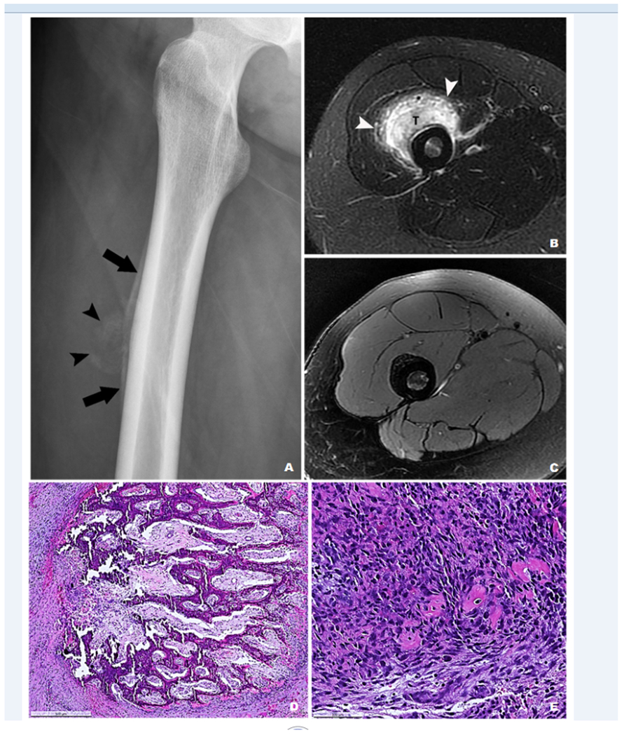
Case 6 – (A) A radiograph showed a juxta-cortical mass with calcifications (arrowheads) and periosteal reaction (arrows). (B) On axial MRI STIR image, the mass (T) was surrounded by edema (arrowheads) and thickened soft tissue in the vastus intermedius muscle. No fluid level was identified. (C) Axial T2-weighted fat-suppressed image of the follow-up MRI 10 months after the open biopsy showed unchanged thick periosteal reaction along the femoral cortex with almost complete resolution of the previously identified high T2 signal, consistent with continued internal healing. (D) An incisional biopsy showed exuberant interconnecting woven bone formation and (E) neighboring areas of fibroblastic proliferation with small foci of osteoid matrix.