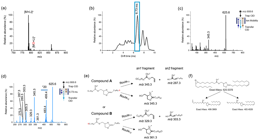
Figure 3.

Positive-ion mode analysis of 20 μM PC(18:1(9Z)/18:1(9Z)) in acetone/water/methanol (75:12.5:12.5) with 5 mM LiOAc added. (a) TENG-MS analysis showing both the [M + Li]+ and epoxidated [MO + Li]+ions. (b) Total ion ATD following selection and trap CID of the [MO + Li]+ ion (m/z 808.6). The blue box represents the region corresponding to the dioxolane fragment ion at m/z 625.6. (c) Averaged TAP mass spectrum across the full ATD. (d) TAP transfer CID spectrum of fragment ions outlined with the blue box in (b). (e) Fragmentation pathways for the two possible dioxolane ions derived from [MO + Li]+. Generic pathways for lipids with other acyl chain lengths are shown in Figure S7. (f) Proposed structures for diagnostic fragments species detected at m/z 499.4 and 483.4 indicating the Δ9 double-bond position.
