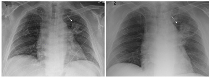
Figure 1.
Chest X-ray showing radiation pneumonitis Image 1—Frontal chest X-ray showing left upper lobe mass (arrow), the patient also had a right internal jugular port placed. Image 2—Post radiation treatment frontal chest X-ray showing increasing alveolar and interstitial opacities in the left upper lobe and in the left lower lobe in a patient suspected of radiation pneumonitis.

