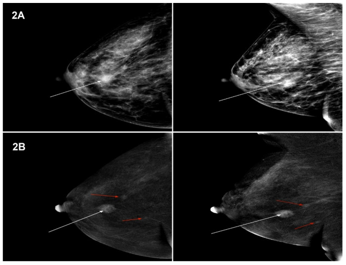
Figure 2.
(A) MG (low-energy images)—tumour in the right breast, in the central part, measuring 16 mm—BI-RADS 6 (white arrow). Projection: CC and MLO, HP-Ca lobulare GIII. (B) CESM (subtraction images)—tumour in the right breast, in the upper external quadrant, measuring 15 mm, enhanced upon contrast injection—BIRADS 6, HP: Ca lobulare GIII (white arrow). Foci of amorphous contrast enhancement of a focal type, measuring 6 mm and 5 mm, at a distance of 12 mm and 26 mm from the tumour—BI-RADS 4 (red arrows). Projection: CC and MLO, HP-Ca lobulare GIII.

