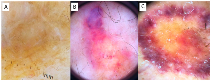
Figure 2.
Dermoscopy. (A) Case 1: a structureless yellow background interspersed with whitish scar-like strikes in Case 1; (B) Case 2: roundish yellow waxy blotches on a hemorrhagic background, interspersed with fine telangiectasias and hemorrhagic spots; (C) Case 3: structureless yellow background interspersed with whitish spots, surrounded by a hemorrhagic halo with elongated serpentine vessels.

