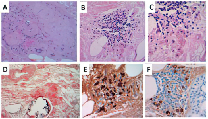
Figure 4.
Histopathological findings of Case 3. (A) presence of a hyaline-like, amorphous eosinophilic material in the dermis surrounding and involving dermal vessels. (HE original staining ×40); (B) A perivascular and interstitial lymphocytic infiltrate (original magnification ×100) with (C) focal plasma cells component (original magnification ×400). (D) Congo red stained positively the amorphous eosinophilic deposits of amyloid in the dermis, surrounding deep dermal vessels (original magnification ×100). Immunohistochemical studies for (E) lambda and (F) kappa light chains showed evidence of lambda light-chain restriction, consistent with a monoclonal plasma cell proliferation.

