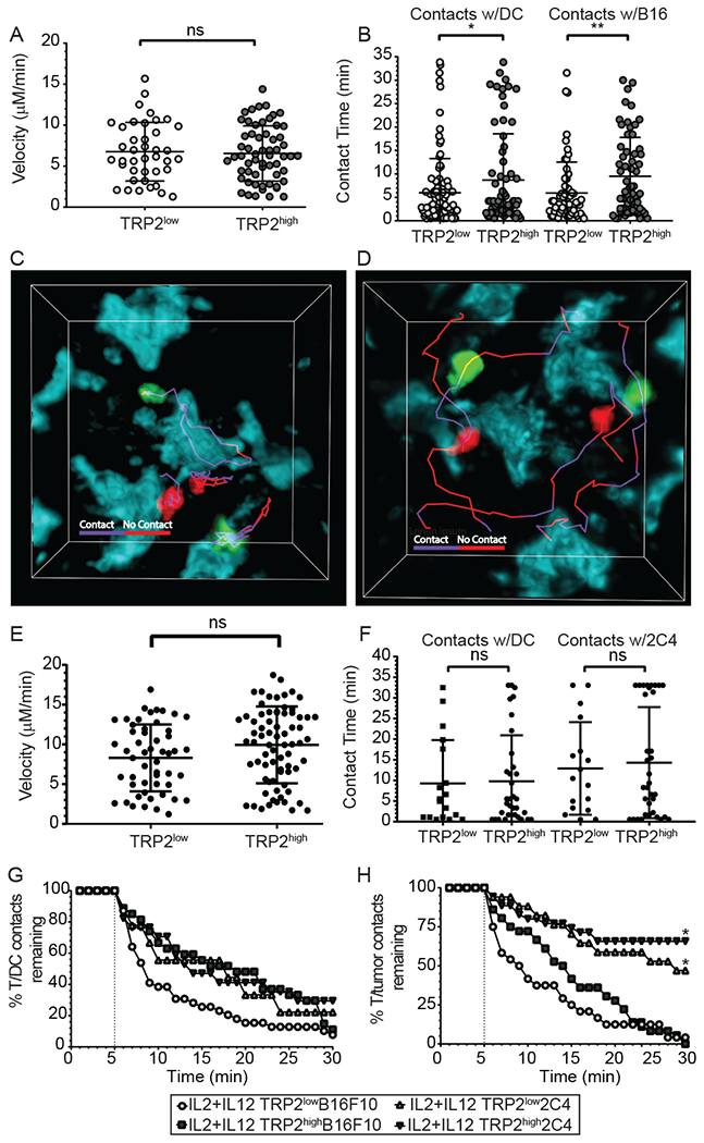
Figure 4: Antigen overexpression restores the ability of IL-12 primed low avidity T-cells to make sustained contacts with tumor targets.

(A-H) CD11c-YFP mice received B16F10-TFP or 2C4 tumors. Ten days after inoculation, mice received 5-10x106 IL-2 plus IL-12 TRP2low and TRP2high T-cells. 72 hours after adoptive T-cell transfer, mice were euthanized and tumors were removed and analyzed by two-photon microscopy. (A, B) T-cells were identified with Brainbow tdTomato (TRP2low) or UBC-GFP (TRP2high) expression and analyzed using Imaris software for average velocity and contact time in B16F10 tumors. (C, D) Representative images showing TRP2low and TRP2high T-cell contacts with tumor cell surfaces using tracks. Purple tracks represent sustained contact with the T-cell and tumor cell, while red tracks represent no-contact between T-cells and tumor targets at the timepoint on the track. Two photon microscopy was performed on ex vivo 2C4 tumors and (E) velocity and (F) contact time with dendritic cells or tumor targets. (G-H) Contact decay was analyzed between TRP2low or TRP2high and (E) YFP+ DCs (F) or TFP+ tumor targets. Contacts lasting less than five minutes were excluded from analysis. Data are from two independent replicates. ns = not statistically different, **=p<0.001, ***=p<0.0001.
