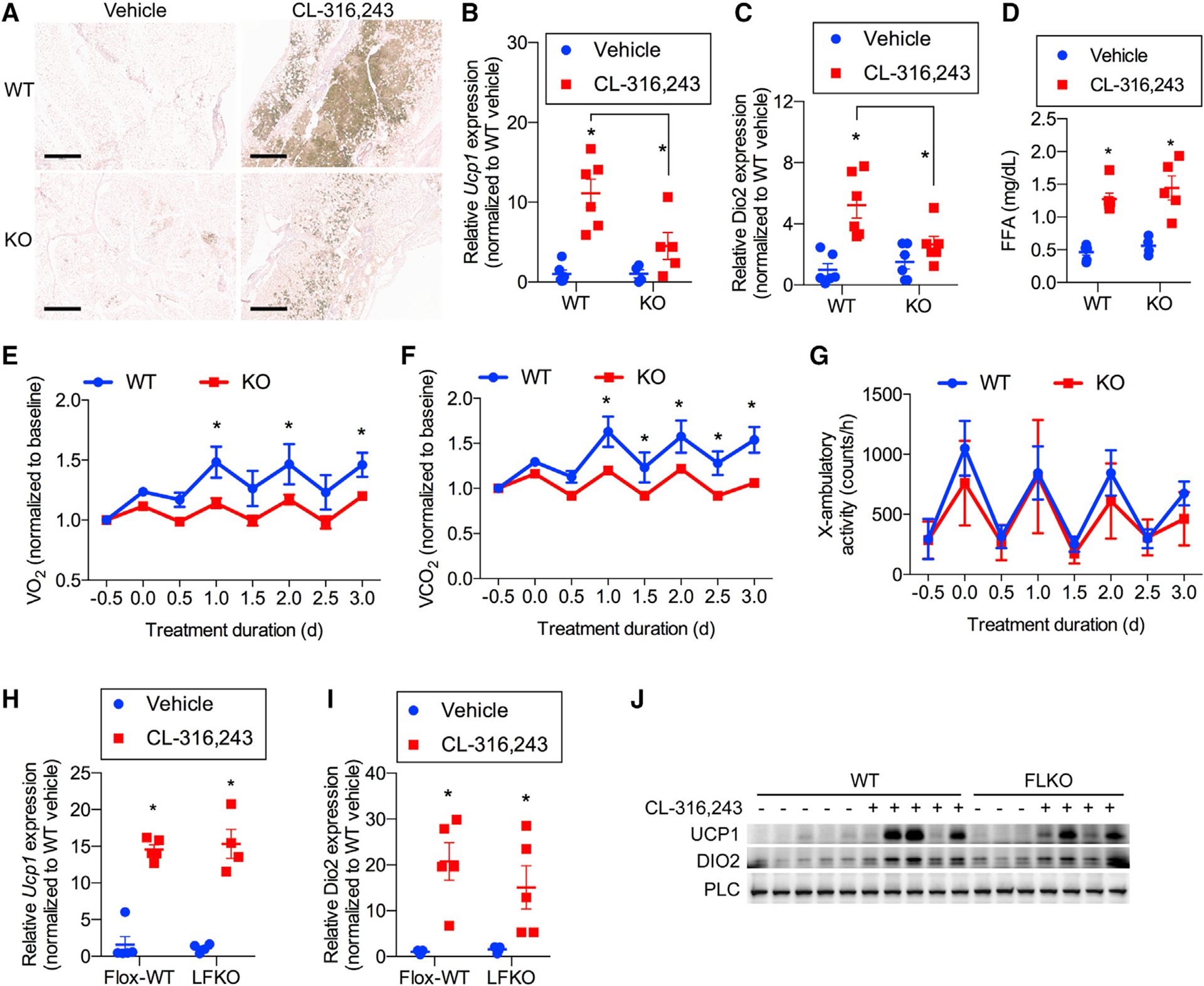
Figure 3. The metabolic benefit of FGF21 is due to its action as an autocrine factor in iWAT.

(A–C) WT and whole-body FGF21-KO mice treated with 1 mg/kg CL or vehicle daily for 7 days. n = 6 mice per genotype per treatment.
(A) Representative immunohistochemistry (IHC) staining of UCP1 in iWAT. Scale bar, 0.5 mM.
(B and C) Ucp1 (B) and Dio2 (C) mRNA expression in iWAT.
(D) Serum free fatty acid (FFA) concentrations 20 min after treatment with 1 mg/kg CL or vehicle in WT and FGF21-KO mice. n = 6 mice per genotype per treatment.
(E–G) Baseline normalized oxygen consumption (E), baseline normalized carbon dioxide production (F), and physical activity (G) as assayed by x axis beam breaks in WT and whole body FGF21-KO mice treated with 1 mg/kg CL daily. n = 6 WT and 7 KO mice.
(H and I) Ucp1 (H) and Dio2 (I) mRNA expression of WT mice and LFKO mice treated with 1 mg/kg CL or vehicle daily for 7 days. n = 5 mice per genotype per treatment.
(J) Western blot analysis of FGF21 protein in iWAT following 7-day treatment with 1 mg/kg CL or vehicle in WT and LFKO mice. n = 5 WT mice per treatment group and 3 vehicle-treated and 4 CL-treated FLKO mice. Data presented as mean ± SEM.*p < 0.05 from Holm-Sidak post hoc analysis after significant two-way ANOVA for vehicle versus CL unless otherwise indicated with a line.
