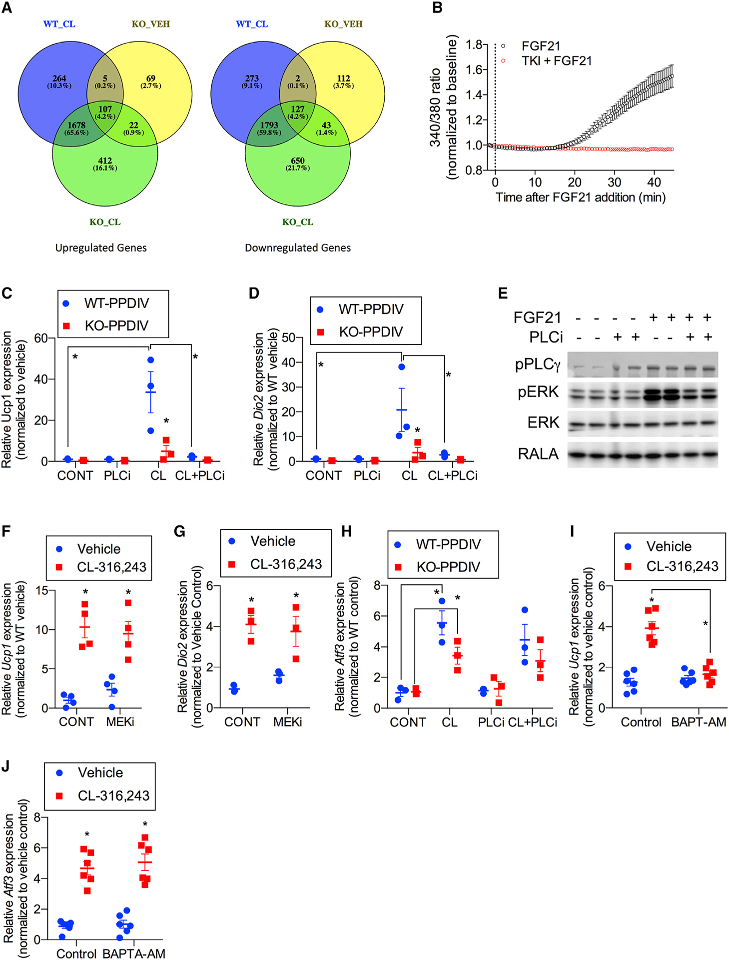
Figure 5. FGF21 provides a second signal that is required for browning of WAT.

Genes involved in calcium signaling are upregulated by CL in a FGF21-dependent manner.
(A) Venn diagrams of differentially expressed genes from RNA-seq analysis of WT and FGF21-KO PPDIVs treated with vehicle or CL (Data S1). n = 3 wells per treatment per genotype.
(B) Effect of FGF21 on calcium mobilization in the absence or presence of 200 nM tyrosine kinase inhibitor (PD173074). PPDIVs were serum starved for 15 h and loaded with fura-2 AM for 15 min in the absence (black trace, n = 63 cells) or presence (red trace, n = 23 cells) of PD173074 before stimulation with FGF21 (200 ng/mL). Curves are pooled from 4 and 2 experiments, respectively. Values are mean ± SEM.
(C and D) Ucp1 (C) and Dio2 (D) mRNA expression in PPDIVs isolated from FGF21-WT mice or FGF21-KO mice; cells were pretreated with 1 μM of PLC inhibitor (U73122) for 30 min, followed by 10 μM CL treatment for 6 h. n = 3 wells per genotype per treatment.
(E) Western blot analysis of PPDIVs pretreated with 1 μM PLC inhibitor (U73122) for 30 min, followed by 100 ng/mL recombinant FGF21 for 10 min. n = 2 wells per treatment. The membrane was blotted against antibodies as indicated.
(F and G) Ucp1 (F) and Dio2 (G) expression in PPDIVs pretreated with 10 μM MEK inhibitorPD0325901 (MEKi) for 30 min, followed by 10 μM CL treatment for 6 h. n = 3–4 wells per cell type per treatment.
(H) Atf3 mRNA expression in PPDIVs isolated from FGF21-WT mice or FGF21-KO mice pretreated with 1 μM of PLC inhibitor (U73122) for 30 min, followed by 10 μM CL treatment for 6 h.
(I and J) Ucp1 (I) and Atf3 (J) mRNA expression in PPDIVs isolated from WT mice. Following differentiation, the cells were pretreated with 10 μM BAPTA-AM for 30 min, followed by 10 μM CL treatment for 6 h. n = 6 wells per cell type per treatment.
Data presented as mean ± SEM.*p < 0.05 from Holm-Sidak post hoc analysis after significant two-way ANOVA for vehicle versus CL unless otherwise indicated with a line.
