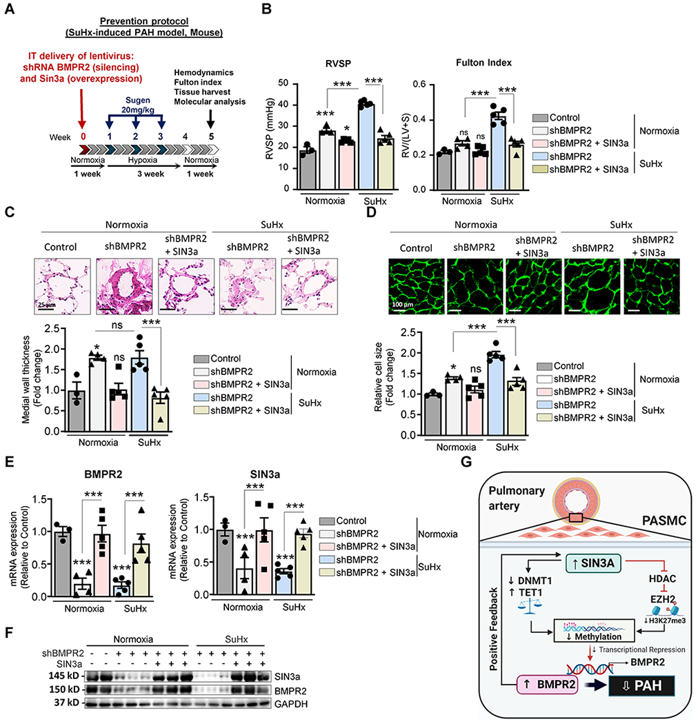
Figure 8. Lentivirus-mediated SIN3a gene transfer attenuated SuHx-induced PAH in shRNA-mediated BMPR2 knockdown mice.

A. Schematic of the experimental design to assess the therapeutic efficacy of SIN3a lentivirus-mediated gene transfer using the SuHx-induced PAH mouse model. Tissues were collected at week 7 for molecular and histology analysis. B. RVSP (left) and Fulton’s index (right) were determined in the indicated conditions. C. Representative hematoxylin and eosin-stained lung sections of the indicated mice. The graph represents the quantification of the medial thickness. D. RV sections were stained with fluorescence-tagged wheat germ agglutinin to measure RV cardiomyocyte cross-sectional area. The graph represents the quantification of the cardiomyocyte size. E. mRNA expression of BMPR2 (left) and SIN3a (right) was assessed by RT-qPCR in the indicated conditions. F. Lung homogenates were analyzed by western blot to assess the protein expression of SIN3a and BMPR2 in control and SuHx-mice treated with shBMPR2 alone or in combination with SIN3a. G. Schematic representation of the molecular mechanisms by which SIN3a inhibits PAH. SIN3a restores BMPR2 expression by a dual mechanism in hPASMC. Our results showed that SIN3a inhibited EZH2 expression and the H3K27me3 contents within the BMPR2 promoter region while decreasing the methylation level of the BMPR2 promoter region by upregulating TET1 and repressing DNMT1 activity, which affects the CTCF binding to the BMPR2 promoter region. Created with BioRender.com. Data are presented as mean ±SEM; ns=not significant, * = p<0.05, *** P < 0.001.
