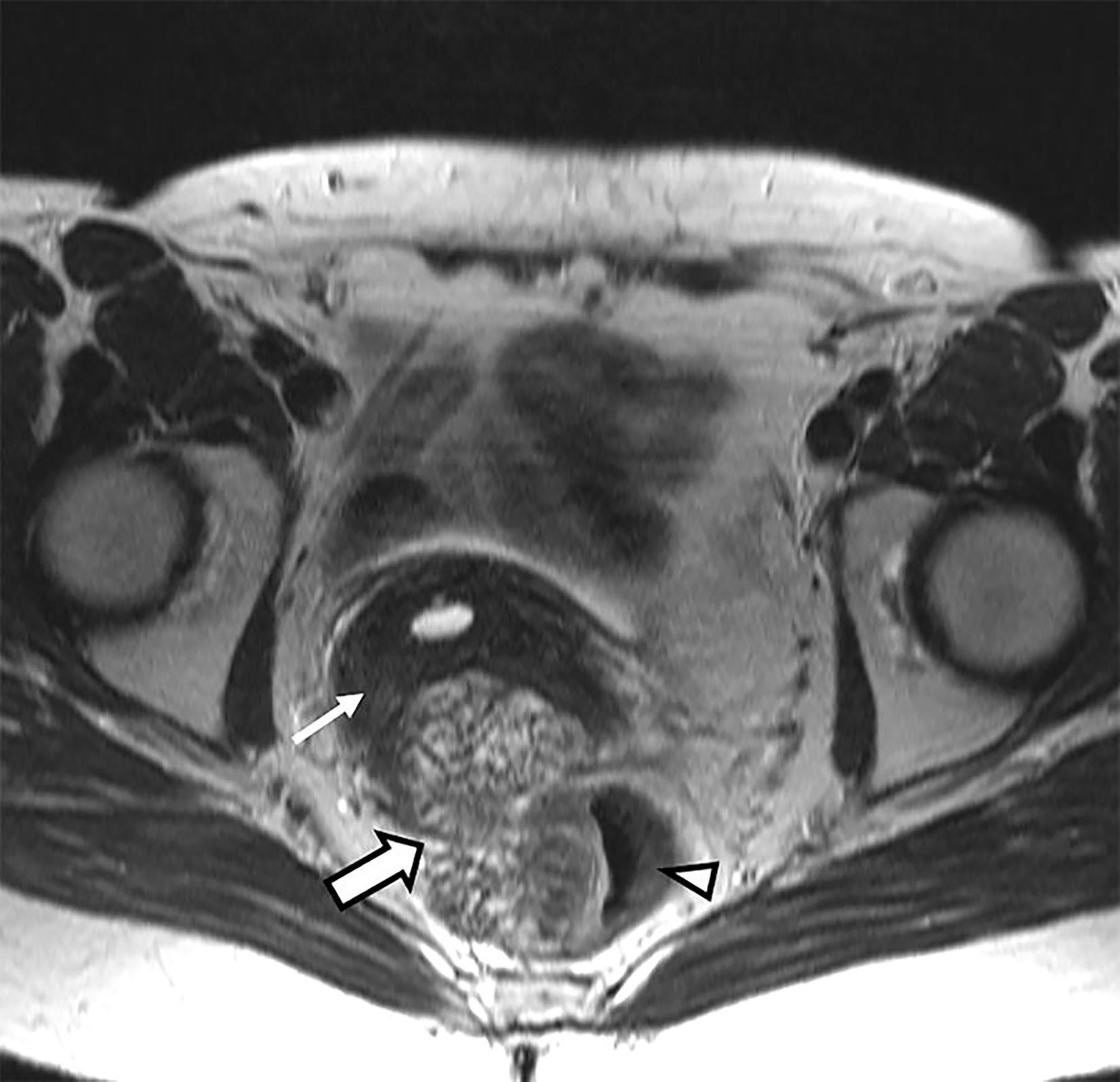
Fig. 1.

Axial T2-weighted image without fat saturation at the level of the cervix. A lobulated, heterogeneously T2-hyperintense mass (thick arrow) is shown to be centered in the cul-de-sac and inseparable from the cervical remnant (thin arrow) and anterior rectum (arrow head); the latter seems to be invaded by the mass
