Fig. 10.

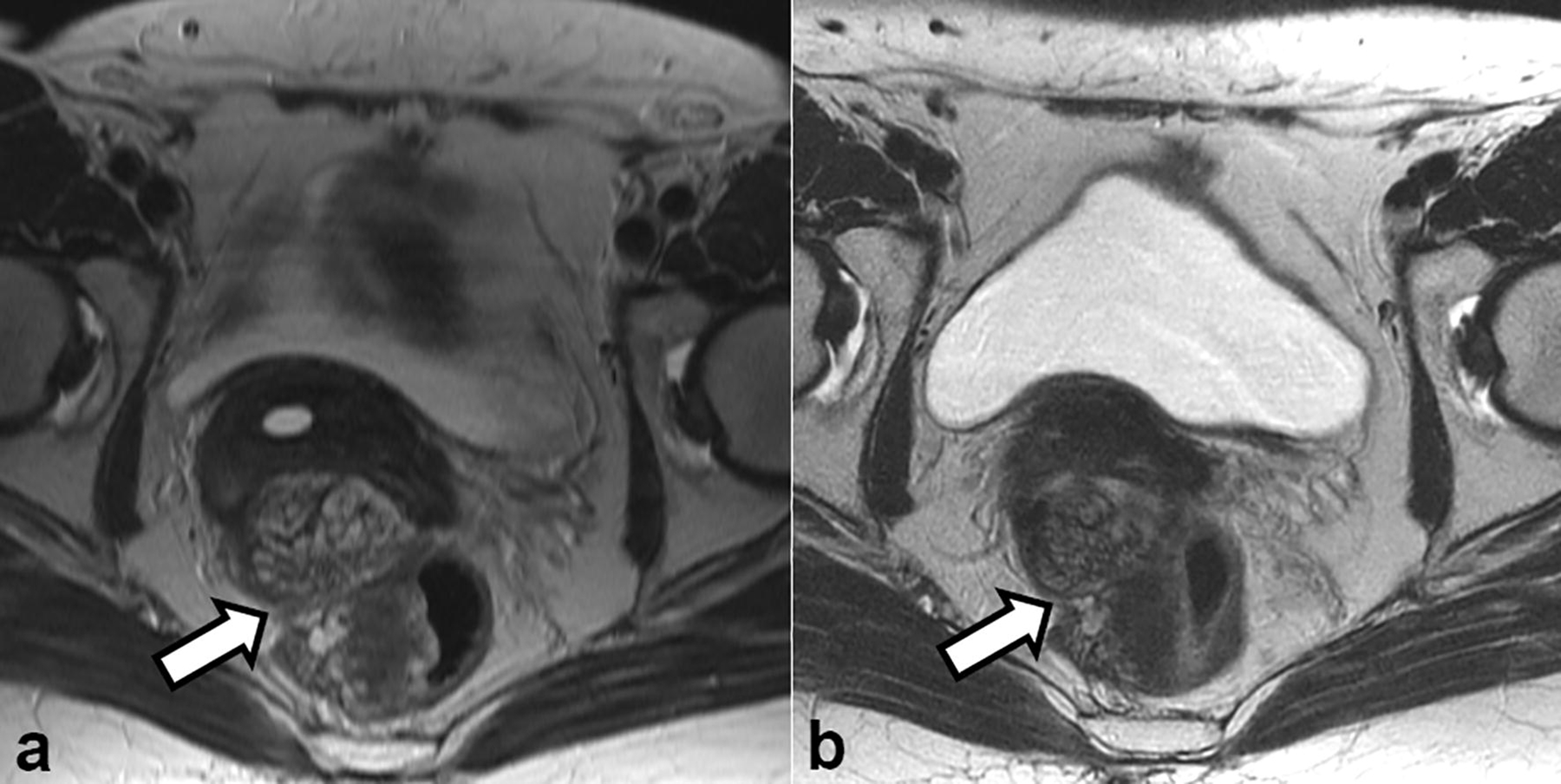
Axial T2-weighted images obtained (a) at initial presentation and (b) 3 months after the discontinuation of hormone replacement therapy showed a slight decrease in the size of the pelvic mass (arrow in a and b)

Axial T2-weighted images obtained (a) at initial presentation and (b) 3 months after the discontinuation of hormone replacement therapy showed a slight decrease in the size of the pelvic mass (arrow in a and b)