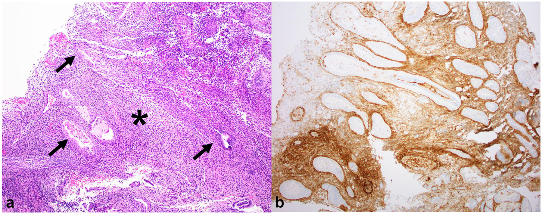
Fig. 9.

(a) Hematoxylin and Eosin (H&E) image of the same biopsy specimen as above at 40× magnification shows simple tubular endometrial glands (thin arrows) lined by columnar cells embedded in stroma with ovoid spindle cells (*) resembling proliferative phase endometrium. (b) On CD10 immunohistochemistry, the stroma stains positive, confirming endometrial-type stroma, while the endometrial glands are negative. The unstained glands are irregularly dispersed with focal back-to-back crowding
