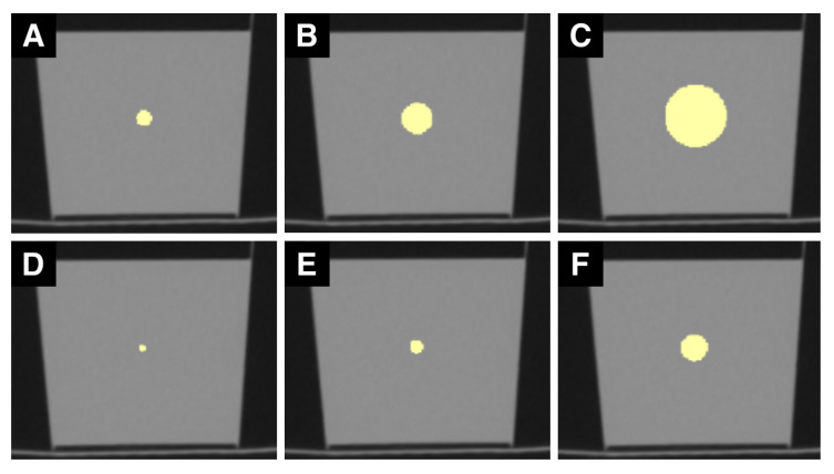
Figure 2.
Sample set of CT ROIs. (A–F) are CT images of the phantom. (A) shows a ROI with a 4 mm diameter, (B) with 8 mm, and (C) with 16 mm. (D) illustrates one slice of the 4-pixel diameter ROI, (E) of the 8-pixel, and (F) of the 16-pixel diameter ROI. mm sized ROIs are generally larger than px sized ROIs.

