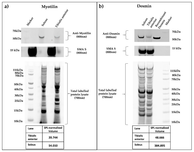
Figure 4.
(a,b) Quantitative Smart Protein Layers (SPL) Western blot analysis of whole murine soleus and tibialis anterior muscle. SPL Western blotting system (NH DyeAGNOSTICS GmbH) was used for the detection of quantitative differences between selected candidate proteins. Total protein was pre-labeled with a red fluorescent fluorophore (700 nm), and a 12.5 kDa green fluorescence-labeled standard protein (SMA S, 800 nm) was spiked in, enabling error correction of differing sample loading and data normalization between experiments. Primary antibodies against myotilin (a), myotilin (E-10), sc-393957), and desmin (b), DAKO, M0760 Clone D33) were visualized by infrared secondary antibodies (IRDye goat anti-mouse 800 nm). SPL system analysis resulted in quantitative protein volumes (SPL normalized volume), determining a higher concentration in the soleus muscle for both proteins.

