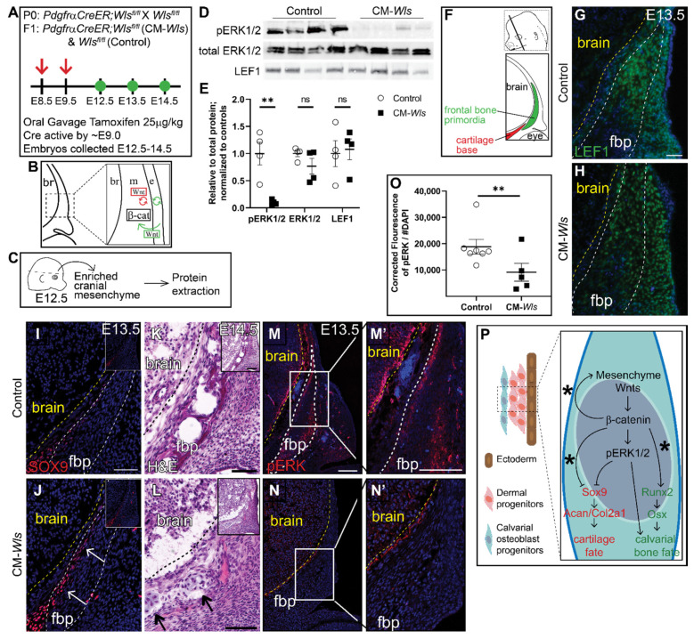
Figure 4.
Mesenchyme Wnts are required for ERK1/2 activation in vivo. (A) Schematic depicting the gavage regimen. (B) Schematic depicting the status of Wnt signaling in the cranial mesenchyme and overlying ectoderm in this mouse model of Wnt signaling deletion. br, brain; m, mesenchyme; e, ectoderm. β-catenin remains intact within the cranial mesenchyme. The ectoderm is able to secrete Wnt ligands and undergo Wnt signaling. The mesenchyme cannot secrete Wnt ligands but can transduce Wnt signaling from the Wnt ligands secreted by the overlying ectoderm. (C) Schematic depicting the work flow and tissue source for the protein analysis. (D,E) Expression of activated ERK1/2 (pERK1/2), total ERK1/2, and LEF1 relative to total protein and normalized to controls in E12.5 control and CM-Wls. n = 4 biological replicates. p(**) = 0.0026. (F) Schematic indicating the plane and orientation of specimens. (G,H) Expression of LEF1 is decreased fbp of PdgfrαCreER;Wlsfl/fl (CM-Wls) relative to controls. (I,J) CM-Wls express ectopic SOX9 in the fbp relative to controls. (K,L) H&E shows ectopic cartilage nodules in CM-Wls but not controls. (M–N’) Immunofluorescence of control and CM-Wls showing diminished pERK1/2 in the CM-Wls relative to the controls. (O) Quantification of pERK1/2 IF by corrected fluorescence using ImageJ. n = 7 controls; 5 mutants. p(**) ≤ 0.01. (P) Summary schematic illustrating the role of Wnts on ERK activation and downstream effect on cell fate decisions in calvarial osteoblast progenitors. (*): Goodnough et al 2016. Arrows point to ectopic cartilage nodule. Yellow line demarcates brain in IF panels; black line demarcates brain in panels K & L. fbp, frontal bone primordia.

