Figure 1.
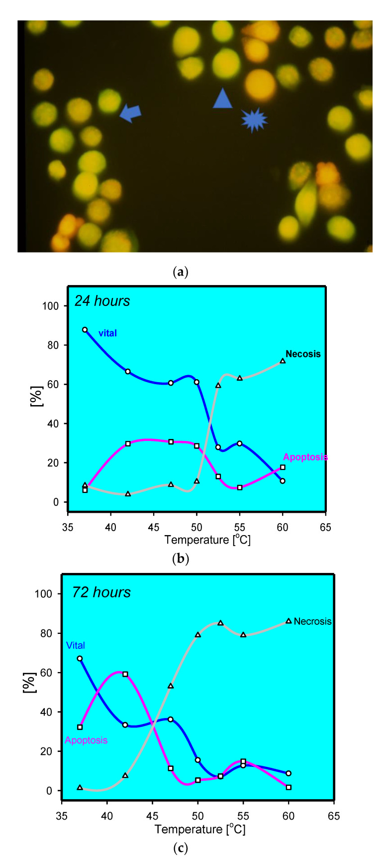
(a) Staining of thermally stress keratinocytes at 47 °C after 72 h. Viable cells have intense green fluorescence (arrow head). Apoptotic cells with reduced green fluorescence and condensed red fluorescence (arrow). Homogenous red fluorescence indicative for necrotic cells (star); (b) percentage of live, apoptotic, and dead keratinocytes after thermal stress for 5 min at the given temperature analysed by acridine orange/ethidiumbromide, indicating a sharp increase in necrotic cells above 50 °C and decrease in viability. Apoptotic keratinocytes were mainly observed at moderate temperatures; (c) percentage of live, apoptotic, and dead keratinocytes after thermal stress for 5 min at the given temperature analysed by acridine orange/ethidiumbromide after 72 h; notice the increase in apoptotic keratinocytes at moderate temperatures.

