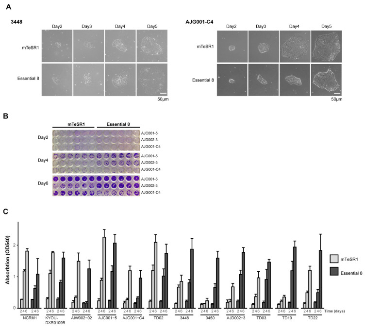
Figure 2.
HiPSC growth and proliferation under different conditions. (A) Representative daily morphology of hiPSCs maintained in mTeSR1 or E8 media. Both cell lines show smooth-edged, tightly packed cells with a large nucleus-to-cytoplasm ratio. AJG001-C4 cells grow slightly better in mTeSR1 media, while 3448 proliferate better in E8. (B) CV assay of representative cell lines grown in mTeSR1 and E8. AJC001-5 grows well in both media. AJG001-C4 grows better in mTeSR1 media, while AJD002-3 cells proliferate best in E8. (C) Quantification of the hiPSCs’ survival and growth in different media. The CV stain was dissolved in methanol and optical density was measured at 540 nm (OD540). The mean and the standard deviation are from six replicates from two independent experiments.

