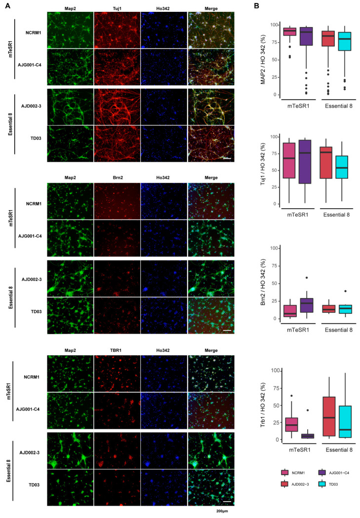
Figure 7.
Immunocytochemistry staining of hiPSC-derived cortical neurons. (A) Representative images of ICC-stained cortical neurons from AJG001-C4, NCRM1, AJD002-3, and TD03 after 4 weeks of neuronal differentiation. Cells were stained with MAP2 and Tuj1 neuronal markers, and cortical neuron markers Brn2 and Tbr1. Nuclei were counterstained with Ho342. (B) Quantification of MAP2-, Tuj1-, Brn2-, or Tbr1-positive cells in (A).

