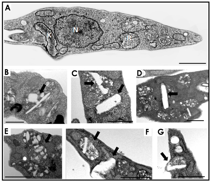
Figure 1.
Effects of lopinavir and nelfinavir on the reservosomes of T. cruzi epimastigotes. (A) Transmission electron microscopy of control parasites revealed the normal morphology of organelles such as nucleus (N), kinetoplast (K) and reservosome (R). The treatment with lopinavir (B–D) and nelfinavir (E–G) induced a huge appearance of lipid inclusions inside the reservosomes (black thick arrows). Bars = 500 nm. The images are a representative set of three independent experiments.

