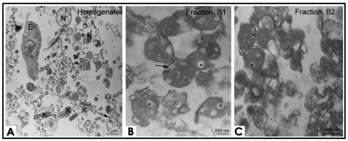
Figure 2.
Ultrastructure of epimastigote cell fractionation by transmission electron microscopy. (A) Total homogenate shows cell debris, reservosomes (R), nuclei (N), whole epimastigote (E), membrane profiles, flagella (black arrow) and kinetoplast (K). Fractions B1 and B2 are enriched in reservosomes, although B1 presents more reservosomes containing lipid inclusions. (B) Cholesterol-rich inclusions are frequently observed in a flattened shape (black arrows) or as a rounded inclusion in the reservosome lumen (asterisk). (C) Reservosomes from Fraction B2 are more electron dense, and lipid inclusions are less abundant (asterisk), suggesting that there are subpopulations of these organelles. The images are a representative set of three independent experiments.

