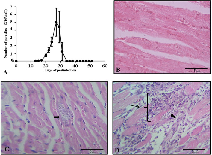
Fig. 1.
Infection and histology. A Parasitemia curve of T. cruzi Ninoa strain in mice over 60 days. B Histological section of normal cardiac tissue of mouse. C Histological section of cardiac tissue is shown during the acute phase; the arrow indicates the presence of amastigote nests with slight presence of mature lymphocytes. D Histological section of cardiac tissue is shown during the indeterminate phase; the arrows indicate amastigote nest, the presence of mature lymphocytes, and a severe inflammatory state with a diagnosis of myocarditis. Histological sections were stained by Hematoxylin–Eosin and observed to 40 X

