
FIGURE 1.
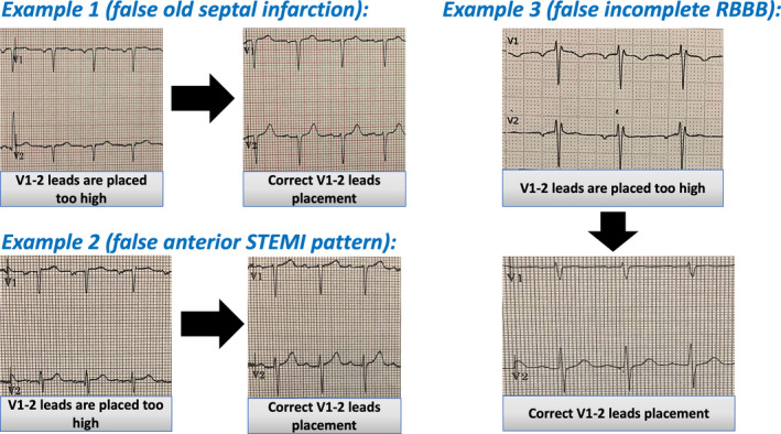
Three real examples of false‐positive ECG findings generated by V1 and V2 leads misplacement. Example 1, incorrect V1‐2 leads placement generated septal Q waves which was misinterpreted as old septal infarction. After correct leads placement, small R waves in V1 and V2 can be seen and septal Q waves disappeared. Example 2, shows Subtle ST elevation in V1 and V2, which was misinterpreted as a possible anterior MI, the ST‐segment elevation in V1 and V2 resolved after correct V1‐2 leads placement. Example 3, shows RsR pattern in V1 and V2 suggestive of incomplete RBBB. Following correct V1‐2 placement, normal RS pattern in V1 and V2 can be seen