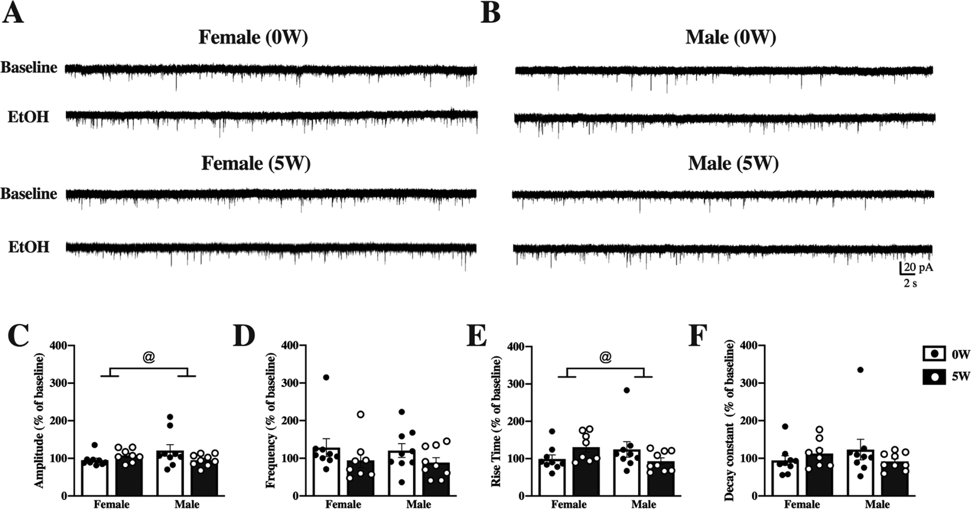
Figure 5. Acute effects of alcohol on DLS neurons differ by sex and drinking history.

Mice were given access to 10% (v/v) alcohol and water in the home cage for 5 weeks (5W, filled bars & white dots) or water alone (0W, open bars & black dots) prior to euthanasia. Whole-cell spontaneous excitatory postsynaptic currents (sEPSCs) were recorded from dorsolateral striatal (DLS) neurons and properties of the currents measured and averaged across a 2-minute time window before (baseline) and after application of 50 mM ethanol (EtOH) to the slices. Representative traces at baseline and after EtOH treatment are shown for neurons from female (A) and male (B) mice with 0W (top) and 5W (bottom) drinking history. (C) Acute EtOH changed sEPSC amplitude differently by sex and drinking history. (D) Frequency of sEPSCs did not differ by sex or drinking history. (E) Acute EtOH increased rise times in 5W but not 0W females and 0W but not 5W males. (F) Decay constants did not differ by sex or drinking history. Histograms represent mean ± SEM baseline-normalized values, calculated as (sEPSC parameter after 50 mM EtOH application)/(sEPSC parameter at baseline). @ p<0.05, sex by drinking history interaction. Number of cells: n=9, 0W female, n=8, 5W female, n=8, 0W male, n=9 D, 5W male; from 4 to 6 mice per group.
