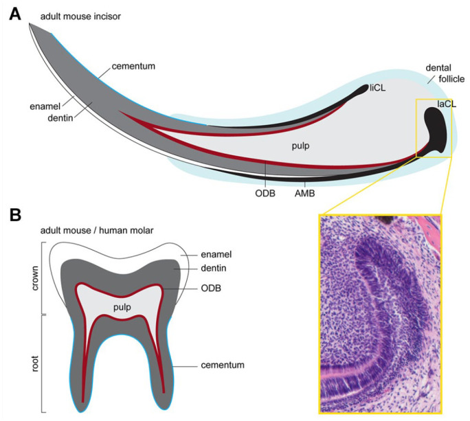
Figure 1.

Mouse tooth structure. (A) Schematics of the continuously gowning mouse incisor. This growth is made possible by the presence of epithelial and mesenchymal stem cells residing at the labial cervical loop (laCL) and the dental pulp, respectively. The epithelial stem cells give rise to enamel secreting ameloblasts (AMBs), while mesenchymal progenitors give rise to odontoblasts (ODBs), which secrete dentin, and also to the cementum-producing cementoblasts and periodontal ligaments. The yellow box shows a hematoxylin and eosin staining of the laCL. Ameloblasts are formed on the labial side, whereas the lingual cervical loop (liCL) is less developed and does not normally produce AMBs. (B) Schematic of the adult mouse molar, which shares many morphological features with adult human teeth. The main difference between mouse and human molars to mouse incisors is that the former have a finite growth phase. Once the molar crown is formed, the progenitors in the epithelial cervical loop are gradually lost, and roots, which anchor the molar to the jawbone, are formed. Pulp also fills the molar and is lined by a layer of ODBs. A layer of enamel covers the dentin at the tooth crown, while the roots are covered with cementum.
