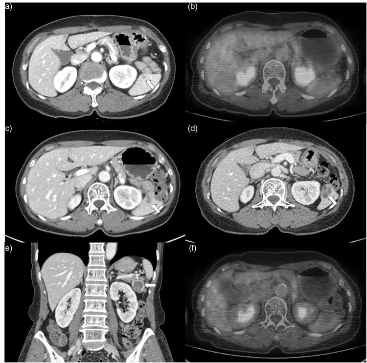
Figure 1.
Splenic mesothelial cyst in a 66-year-old woman with ascending colon cancer. Contrast-enhanced abdominopelvic computed tomography (CT) at first postoperative follow-up shows a 3-mm sized, ill-defined hypodense lesion (arrow) at the spleen (a). There was no significant 18F-fluorodeoxyglucose (FDG) uptake and no other remarkable findings on a positron emission tomography CT (PET-CT) scan (b). After the 3-year follow-up, the lesion had grown to 1.2 × 0.7 cm and had ill-defined, lobulated and heterogeneously enhancing features (arrow) on a CT scan (c). The lesion progressively enlarged and measured 2.3 × 2.5 cm 3 years later (6 years after the surgery). The characteristics had changed to being relatively well-defined, multilocular and low attenuation with enhancing septa (arrow) on axial (d) and coronal (e) CT images. There was no FDG uptake at the splenic lesion on a repeated PET-CT scan (f). The colour version of this figure is available at: http://imr.sagepub.com.

