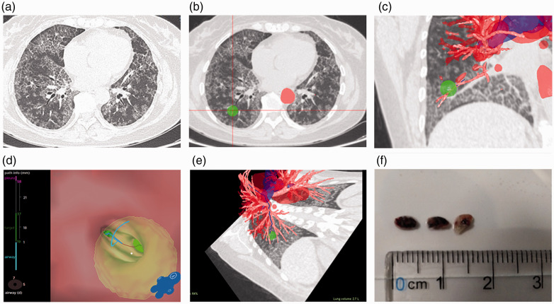
Figure 1.
Different images of the region of interest. (a) Computed tomography images for cryobiopsy show diffuse pavers in both lungs of Patient 6. (b) The region of interest is marked. (c) Coronal view of the region of interest. (d) Distance from the bronchial orifice to the region of interest is measured. (e) Path of endotracheal endoscope. (f) Three lung tissue specimens successfully extracted under the Archimedes Navigation System.

