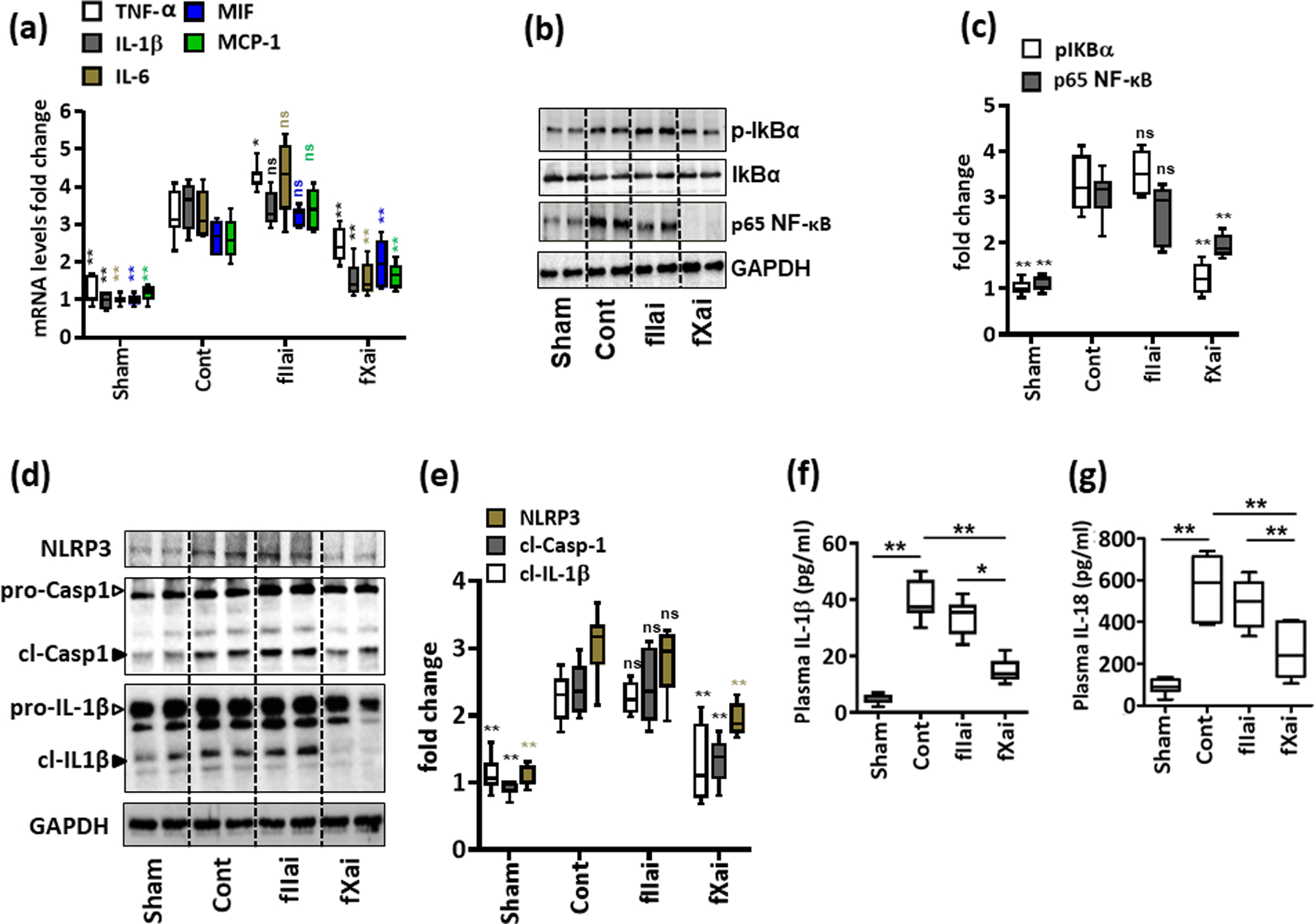
Fig. 3: fXai, but not fIIai, restricts inflammation and NF-κβ activation following myocardial IRI.

a: fIIai- and fXai differentially regulate inflammation in infarcted myocardial tissue. mRNA expressions (quantitative RT-PCR) of pro-inflammatory cytokines interleukin-6 (IL-6), IL-1β, tumor necrosis factor-α (TNF-α), macrophage migration inhibitory factor (MIF) and monocyte chemoattractant protein-1 (MCP-1) were induced following myocardial IRI. fXai but not fIIai inhibits mRNA expression of IL-6, IL-1β, TNF-α, MIF and MCP-1. Box-plots summarizing data of qRT-PCR. GAPDH was used for normalization.
b,c: fXai reduces NF-κB pathway activation following myocardial IRI. Representative immunoblots (b, GAPDH as loading control) and box-plot summarizing data for phosphorylated levels of IκBα and p65 NF-κB (c).
d-g: Treatment of mice with fXai restricts markers of inflammasome activation following myocardial IRI. Representative immunoblots showing cardiac NLRP3 expression and cleaved caspase- 1 (cl-Casp1) and cleaved IL-1β (cl-IL-1β), loading control: GAPDH (d). Arrowheads indicate inactive (white arrowheads) and active (black arrowheads) form of caspase-1 or IL-1β. The active form was quantified. Box-plots summarizing results (e). Box-plots summarizing plasma levels of IL-1β (f) and IL-18 (g).
Mice without (Cont) or with fXai or with fIIai pretreatment. a, c, e: n=8; f, g: n=10 for each group; *P<0.05,**P<0.01, ns: non-significant (compared to Cont), a, c, e-g: ANOVA comparing control to other groups.
