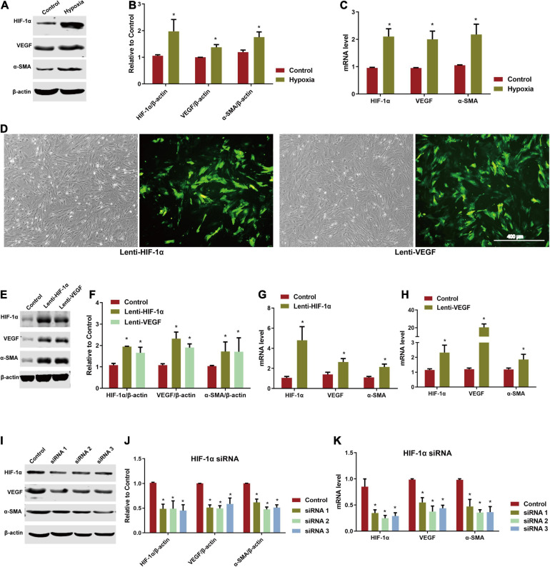
FIGURE 3.
Involvement of HIF-1α/VEGF pathway in directed induction of endothelial differentiation of ADSCs. (A) Expression of HIF-1α, VEGF, and α-SMA of hypoxia-induced ADSCs using western blotting. (B) Analysis of HIF-1α, VEGF, and α-SMA by western blotting. *P < 0.05 vs. the control (normoxic) group. (C) mRNA level of hypoxia-induced HIF-1α, VEGF, and α-SMA in ADSCs by RT-qPCR testing. *P < 0.05 vs. the control (normoxic) group. (D) ADSCs transfected by lenti-HIF-1α and lenti-VEGF for 72 h (Scale bar = 400 μm). (E,F) Expression and analysis of HIF-1α, VEGF, and α-SMA of lenti-HIF-1α and lenti-VEGF transfected ADSCs by western blotting. *P < 0.05 vs. the control (blank vector) group. (G,H) mRNA levels of HIF-1α, VEGF, and α-SMA of lenti-HIF-1α and lenti-VEGF transfected ADSCs by RT-qPCR testing. *P < 0.05 vs. the control (blank vector) group. (I,J) Expression and analysis of HIF-1α, VEGF, and α-SMA of ADSCs knocked down by HIF-1α siRNA detected by western blotting. *P < 0.05 vs. the control (negative) group. (K) mRNA levels of HIF-1α, VEGF, and α-SMA of ADSCs knocked down by HIF-1α siRNA detected using RT-qPCR. *P < 0.05 vs. the control (negative) group.

