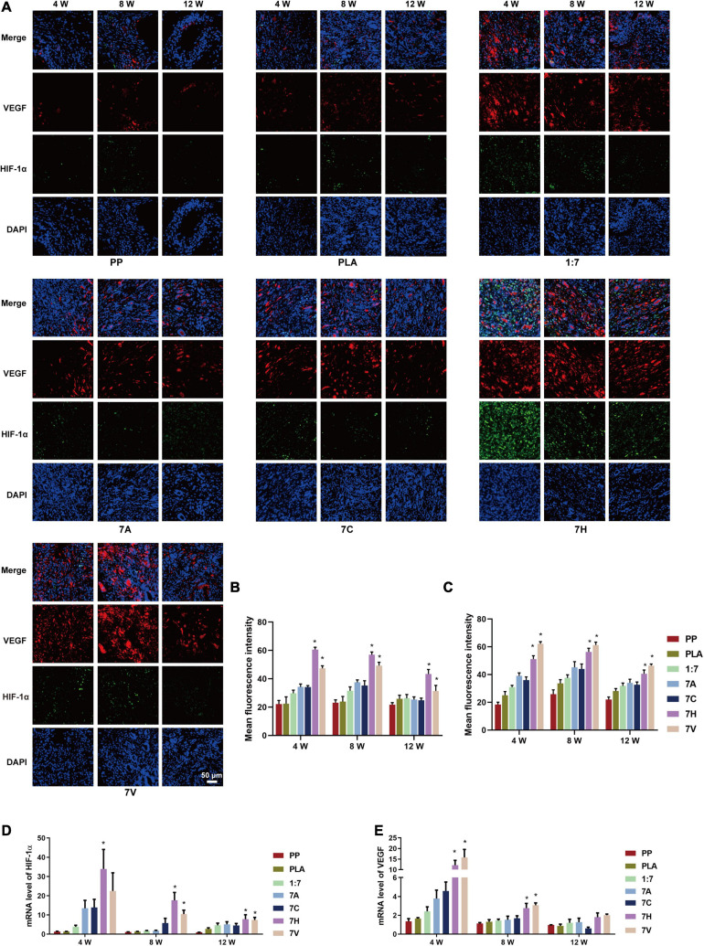
FIGURE 7.
Expression of HIF-1α/VEGF in regenerative tissue of abdominal wall defect model. (A) Expression of HIF-1α and VEGF in regenerative tissue detected by immunofluorescent staining (green: HIF-1α, red: VEGF, blue: DAPI) (Scale bar = 50 μm). (B) Mean fluorescence intensity of HIF-1α. (C) Mean fluorescence intensity of VEGF. (D,E) mRNA levels of HIF-1α and VEGF in regenerative tissue detected by RT-PCR testing. *P < 0.05 vs. the PP, PLA, 1:7, 7A, and 7C groups.

