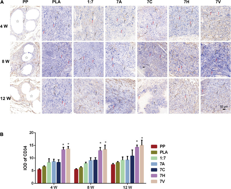
FIGURE 8.
CD34 expression in regenerative tissue detected and analyzed by immunohistochemistry. (A) Microscopic morphology of regenerative tissue (Scale bar = 50 μm). Stars indicate PP mesh fibers. Black arrows indicate vessel lumen and CD34 expression in regenerative tissue. Red arrows indicate foreign body giant cells in regenerative tissue. (B) IOD was used to analyze the expression of CD34. *P < 0.05 vs. the PP, PLA, 1:7, 7A, and 7C groups.

