. 2021 Jun 28;12(28):9839–9850. doi: 10.1039/d1sc02991f

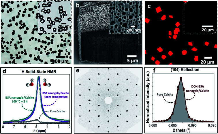
Fig. 3. (a) Optical image of the BSA nanogels occluded in calcite single crystals. The inset shows calcite crystals precipitated in the presence of DOX only. The colorless and transparent crystals indicate that DOX molecules do not incorporate in calcite on their own. (b) FIB-SEM micrograph showing the uniform incorporation of the DOX–BSA nanogels in calcite. (c) Fluorescence image of the DOX–BSA nanogels occluded in calcite (under UV irradiation at 365 nm). Inset is the non-fluorescent BSA nanogel/calcite composites under UV light. The highly fluorescent DOX-nanogel/calcite composites provide evidence of the efficient incorporation of the drug within the hybrid crystals. (d) 1H solid-state NMR of the composites revealing that they are highly hydrated, even after annealing at 100 °C for 2 h. (e) Single-crystal XRD of the hybrid showing that incorporation of the nanogels does not alter the single crystallinity of the calcite host. (f) HR-PXRD showing the main (104) reflection of pure calcite and DOX–BSA nanogels incorporated in calcite.

Fig. 3