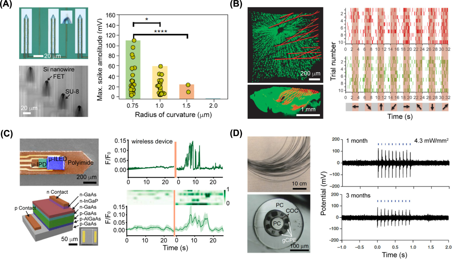
Figure 3. Soft–Hard Composites Can Sense in Neuronal Signals.

(A) Ultra-small 3D transistor probes for intracellular recording. (Top left) Optical image of 3D transistor probes with magnified view (inset) showing that the U-shaped nanowire is transferred onto the device tip. (Bottom left) Optical image of the bend-up device array in water. (Right) Plot of maximum spike amplitude obtained from action potential recordings in dorsal root ganglion neurons using the ultra-small 3D transistor probes. The P values of the statistical studies were obtained using Student’s t-test. *P < 0.1, ****P < 0.0001. Adapted, with permission, from [30]. (B) Mesh electronics for chronic recording in retina from awake mice at the single-neuron level. (Top left) Ex vivo imaging of the interface between the retinal ganglion cells (green) and injected mesh electronics (red) on day 7 after injection (top view). (Bottom left) Side view image of the interface. (Right) Plots showing the firing events of two neurons (red and green) in response to grating stimulations on day 7 after injection. Shaded regions (orange) correspond to times when gratings stimulations were performed. Adapted, with permission, from [53]. (C) Wireless optoelectronic photometer neuron recording in deep brain. (Top left) Colorized scanning-electron-microscopy (SEM) image of the probe. (Bottom left) Schematic of a GaAs μ-IPD with a representative SEM image of μ-IPD in the right corner. (Top right) Plot of fluorescence changes before/after animal was shocked with wireless optoelectronic photometers. (Bottom right) Heatmap of signals (eight trails) recorded before/after animal was shocked. Heatmap is aligned with plotted trace. Adapted, with permission, from [62]. (D) Flexible polymer fibers capable of collocated neural recordings. (Top left) Photograph of a bundle of multimodal fibers (before etching of the sacrificial polycarbonate cladding). (Bottom left) Cross-sectional image of the multimodal fiber. (Right) Electrophysiological recording plots of optically evoked potentials in the medial prefrontal cortex of wild type mice performed at 1 month and 3 months after the one-step implantation and transfection surgery. Adapted, with permission, from [64]. Abbreviations: COC, Cyclic olefin copolymer; FET, field-effect transistor; gCPE, conductive polyethylene (CPE) and 5% graphite; PC, polycarbonate.
