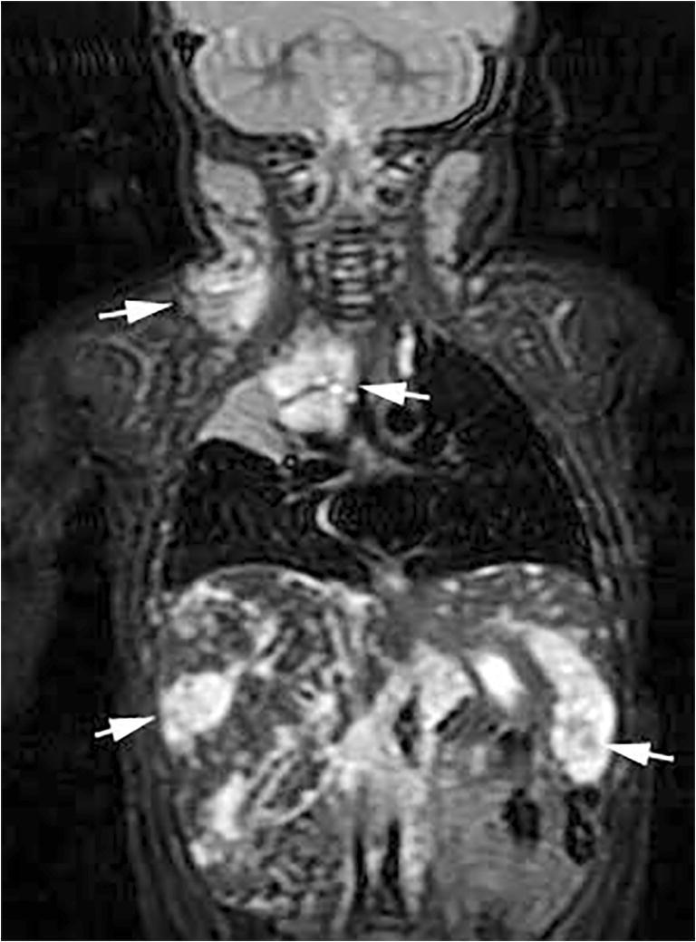
Fig. 1.

Kaposiform lymphangiomatosis at 7 months old. Coronal fat-suppressed T2-weighted MR image shows multiple T2 hyperintense lesions (arrows) in the neck, mediastinum, liver and spleen. These lesions did not enhance after contrast administration (not shown) and were compatible with lymphatic malformations
