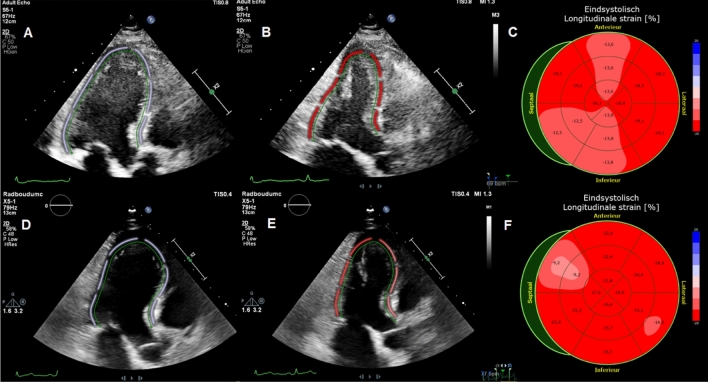
Fig. 2.
Typical case. Typical case of a patient with abnormal global longitudinal strain during hospitalization (− 16.1%) with normal left ventricular ejection fraction (54%) and normal global longitudinal strain (− 18.8%) and left ventricular ejection fraction (54%) at 4 months follow-up. A: apical 3 chamber with end diastolic tracing of left ventricle. B: apical 3 chamber with end systolic tracing of left ventricle. C: bull’s eye plot of global longitudinal strain. D: apical 3 chamber with end diastolic tracing of left ventricle. E: apical 3 chamber with end systolic tracing of left ventricle. F: bull’s eye plot of global longitudinal strain. Image A–C are during hospitalization and D–F at 4 months follow-up

