Figure 6.
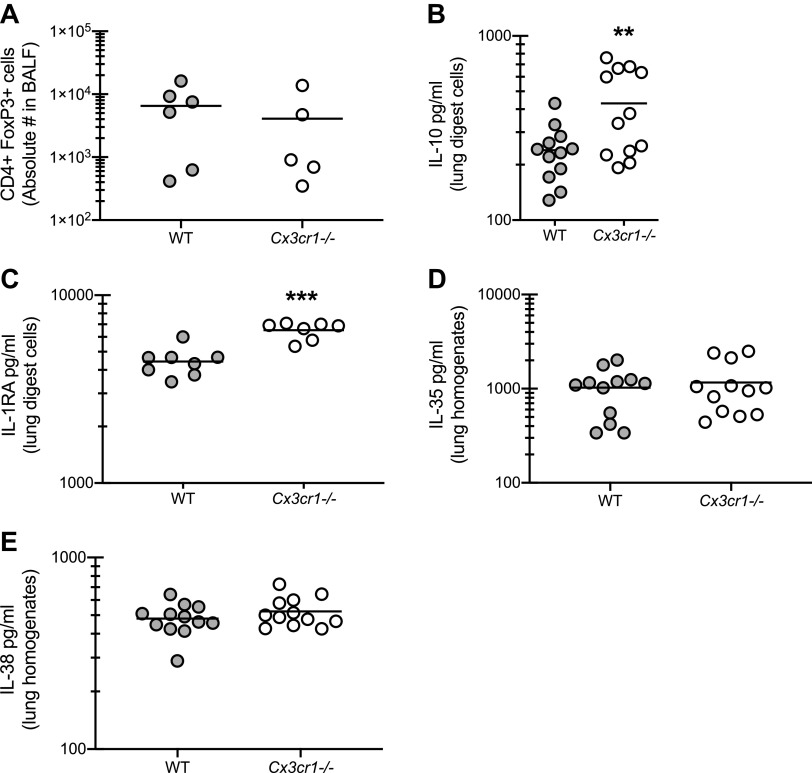
Anti-inflammatory mechanisms are not impaired in the absence of CX3CR1 signaling during fungal-associated allergic airway inflammation. A: C57BL/6 [wild-type (WT)] and CX3CR1-deficient (Cx3cr1-/-) mice were chronically exposed to Aspergillus fumigatus as described in materials and methods. Twenty-four hours after the last organism challenge, lung cells were isolated by bronchoalveolar lavage, enumerated, Fc-blocked, stained with a live/dead staining kit, and stained for T-regulatory cells (CD4+, FoxP3+). A illustrates cumulative data from 2–3 independent studies (n = 2–3 mice per group per study). Twenty-four hours after last challenge, right lungs were collected and enzymatically digested and unfractionated lung cells were cocultured with A. fumigatus conidia for 24 h at a cell to organism ratio of 1:1. B and C: IL-10 (B) and IL-1RA (C) levels were quantified in lung digest cell culture supernatants by MilliPlex or ELISA. B and C illustrate cumulative data from 3–4 independent studies (n = 3–5 mice per group per study). **P < 0.01 and ***P < 0.001 (two-tailed Student’s t test). D and E: 24 h after last challenge, the left lungs were collected and homogenized and IL-35 (D) and IL-38 (E) levels were quantified in clarified lung homogenates by ELISA. D and E illustrate cumulative data from 3–4 independent studies (n = 3–5 mice per group per study). **P < 0.01 (two-tailed Student’s t test).

