Figure 1.
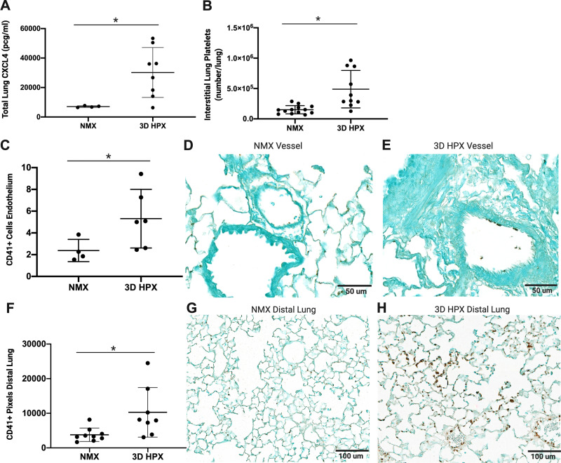
Platelets are increased in the lungs of mice following hypoxia. A: CXCL4 levels in mouse whole lung homogenates, *P < 0.01 by t test (n = number of rats), normoxia (NMX): n = 4 male; 3D hypoxia (HPX): n = 7 male, 1 female. B: lung interstitial platelets in mouse whole lung homogenates, **P < 0.001 by t test, NMX: n = 8 male, 5 female; 3D HPX: n = 6 male, 4 female. CD41+ cells on the endothelium of pulmonary arterioles between 50 and 200 µm (C) NMX, ×40 magnification (D), and HPX, ×40 magnification (E); scale bars = 50 µm, *P < 0.05 by t test; NMX: n = 3 male, 1 female; HPX: n = 3 male, 3 female. Analysis and representative CD41 staining in mouse distal lung sections (F) NMX, ×20 magnification (G), and HPX, ×20 magnification (H); scale bars = 100 µm, *P < 0.05 by t test; NMX: n = 6 male, 3 female; HPX: n = 5 male, 3 female.

