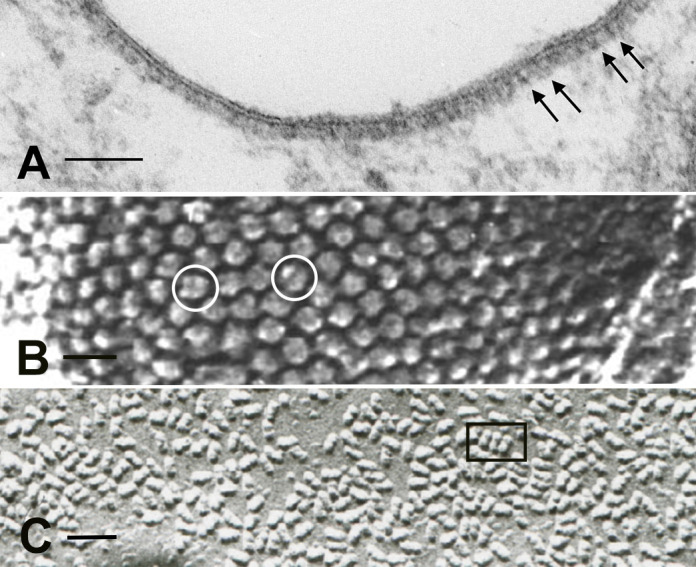
Figure 3.

Microscopy of the V-ATPase in proton-secreting cells. The large cytoplasmic V1 domain of the V-ATPase is clearly detectable as an electron dense array of coating material attached to the underside of the lipid bilayer in a kidney intercalated cell apical plasma membrane (A; arrows; bar = 0.1 µm). The rapid-freeze, deep etching procedure reveals the dense, hexagonally packed arrays of V1 domain stud-like projections attached to a vesicle in a proton-secreting cell from toad urinary bladder (B; circles; bar = 25 nm). The freeze fracture technique, exposing the internal domain of split-open lipid bilayers, reveals numerous so-called rod-shaped or dumbbell-shaped (e. g., inside the rectangle) intramembranous particles in V-ATPase-rich membranes: this image is from a kidney intercalated cell (C; bar = 50 nm).
