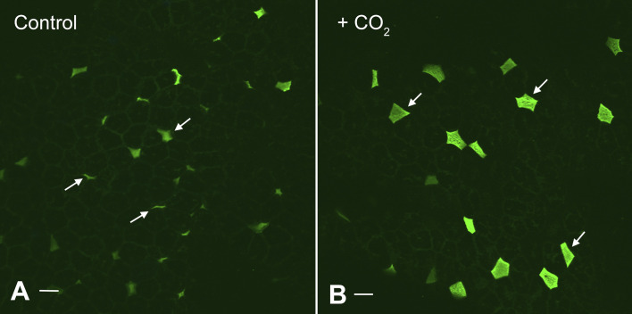
Figure 6.
Effect of CO2 exposure on expression of V-ATPase and apical membrane area of proton-secreting cells in the turtle urinary bladder. Image shows a whole mount view of the apical epithelial surface of a turtle urinary bladder stained with antibodies against the “A” subunit of the V-ATPase, followed by secondary donkey anti-rabbit Ig coupled to Alexafluor 488. The V-ATPase-rich cells (arrows) show a remarkable increase in surface area between the control (A) and tissues acutely exposed to basolateral CO2 (B), indicating that CO2 rapidly activates these cells. Tissue was kindly provided by Dr. John Schwartz, Boston University Medical Center, Boston who exposed bladders to CO2 as part of the annual “Origins of Renal Physiology” course at Mount Desert Island Biological Laboratories, Bar Harbor, ME. Bar = 10 µm.

