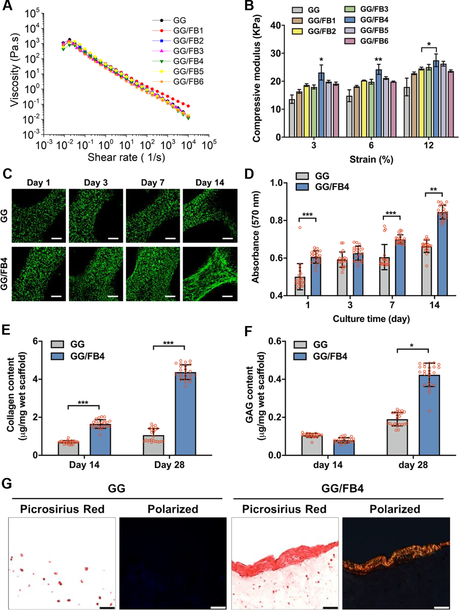
Figure 2.

Material characterization and in vitro biological evaluation of GG/FB bioinks. (A) The viscosity of GG/FB bioinks with different concentrations of FB before crosslinking and (B) compressive elastic modulus of GG/FB hydrogel constructs after crosslinking. (C) Live/dead stained images of the printed pMCs in GG alone and GG/FB4 constructs showing viability at 1, 3, 7, and 14 days in culture, where live cells were stained in green and dead cells in red (scale bar: 200 μm). (D) Metabolic activity of the printed pMCs in GG and GG/FB4 constructs at 1, 3, 7, and 14 days in culture as confirmed by AlamarBlue assay. Quantification of (E) collagen and (F) GAG production in GG alone and GG/FB4 constructs at 14 and 28 days in culture. (G) Non-polarized (left) and polarized (right) Picrosirius Red stained images of the bioprinted cell-laden GG and GG/FB4 constructs after 28 days of culture (scale bar: 50 μm). Statistical significant differences were represented by *p < 0.5, **p < 0.01, and ***p < 0.001. GG alone without FB was used as control.
