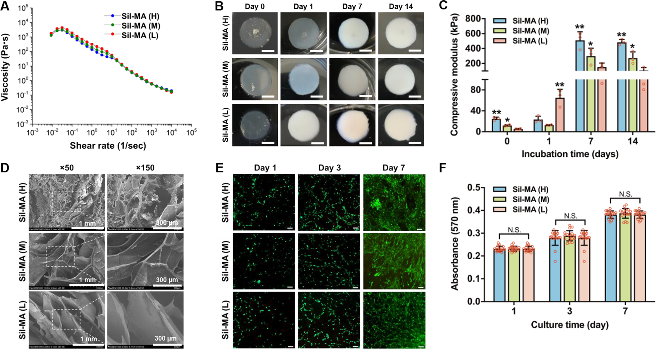
Figure 3.

Material characterization and in vitro biological evaluation of Sil-MA bioinks. (A) The viscosity of Sil-MA bioinks with different methacrylate degrees. (B) The conformational change occurred during incubation at 0, 1, 7, and 14 days of incubation. (C) Compressive elastic modulus of Sil-MA constructs at 0, 1, 7, and 14 days in culture (*p < 0.05 compared with Sil-MA (L) at 0, 7, and 14 days; *p < 0.05 compared with Sil-MA (M) at 1 day and **p < 0.05 compared with others). (D) SEM images of Sil-MA constructs with different methacrylate degrees. (E) Live/dead stained images of pMCs seeded Sil-MA constructs at 1, 3, and 7 days in culture, where live cells were stained in green and dead cells in red (scale bar: 200 μm). (F) Metabolic activity of pMCs seeded in Sil-MA constructs at 1, 3, and 7 days in culture as confirmed by AlamarBlue assay (N.S.: no significant).
