Figure 4.
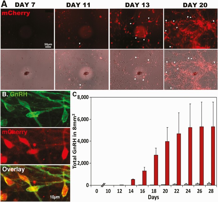
Fluorescent mCherry-labeled GnRH neurons derived from hESC allowed us to clarify the time course of GnRH neurogeneration. Using the CRISPR/Cas9 gene editing system, hESCs were modified such that mCherry is expressed when the GnRH gene turns on. (A) Live cell imaging (top row) and phase contrast imaging (second raw) were taken on days 7, 11, 13, and 20 of the FGF8 exposure at the same slide position, as indicated by a black marker with a white shadow circle. Note that because of the migratory nature of GnRH neurons, GnRH neurons seen one day are not visible in the same location in the later days. (B) An overlay microphotograph (bottom panel) indicates that mCherry expressing cells (red, middle panel) are also GnRH peptide positive (green, top panel). Note that while the peptide staining is seen in the cell body as well as in neurites, mRNA expression is limited to the cell body. (C) GnRH neurons counted in an 8-mm2 area showed that GnRH neurons are seen as early as days 11 and 12 of FGF8 exposure, and the number of GnRH expressing cells progressively increased between days 12 and 24, reaching the plateau. Red bars indicate cultures exposed to FGF8, whereas gray bars indicate cultures exposed to NDM alone. n = 4/group. For statistical analyses, see Supplementary Table 4 in (26).

