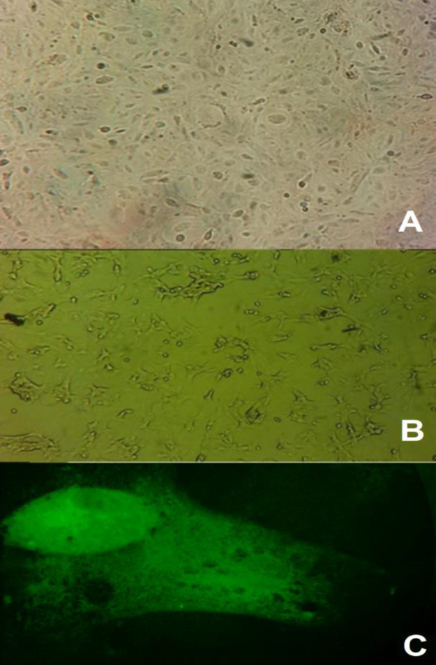
Fig. 1.
Bovine endometrial epithelial cells cultures. (A) Normal appearance of epithelial-like cells after 7th passaging (×20), (B) Appearance of epithelial cells of bovine endometrium at 10th passaging (×20), and (C) Immunofluorescence assay of bovine endometrial cells stained with Alexa 488 (green) showed the presence of Cytokeratin 18 (×40)

