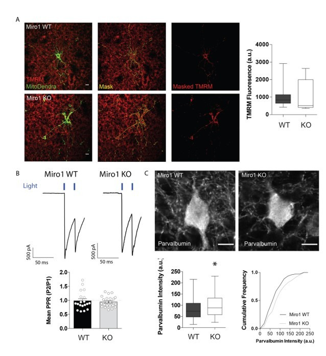
Author response image 1. Parvalbumin levels seem to be increased in the Miro1 KO while mitochondrial TMRM fluorescence and synaptic facilitation are unaffected by the absence of Miro1.

(A) Loss of Miro1 does not affect mitochondrial TMRM uptake in parvalbumin interneurons. Representative confocal images from control and cKO organotypic brain slices that were bulk loaded with TMRM. The MitoDendra signal was used as a mask to isolate the TMRM signal emerging from mitochondria in parvalbumin interneurons. Scale Bar = 10 μm. Boxplot shows the quantification for the mean TMRM fluorescence in the cell (nWT = 28 neurons from 6 slices from 3 animals, nKO = 21 neurons from 4 slices from 2 animals). (B) Loss of Miro1 does not alter short-term facilitation. Example responses from control and Miro1 KO cells in the hippocampus. The bar chart shows the quantification for the mean paired-pulse ratio (PPR) of the second response divided by the first response to light and the error bars represent the standard error of the mean (nWT = 19, nKO = 22 recordings from 4 animals). (C) Loss of Miro1 increases parvalbumin levels in the hippocampus. Boxplot and cumulative frequency distribution of the parvalbumin fluorescent intensity signal (nWT = 79 neurons from 10 slices from 3 animals, nKO = 73 neurons from 8 slices from 3 animals).
