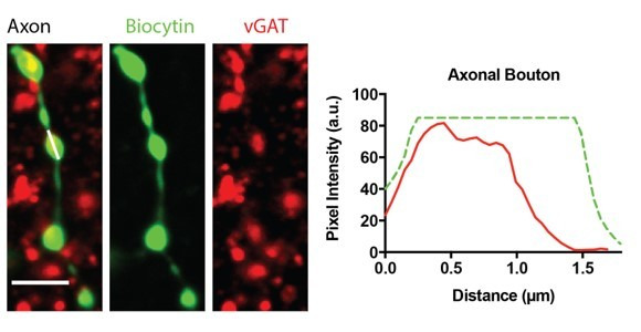
Author response image 2. Biocytin-filled boutons on the PV+ interneuron axon represent inhibitory pre-synaptic terminals.

Example max-projected confocal images of biocytin-filled axon. Scale Bar = 3 μm. The graphs represent the fluorescence signal of vGAT (red) from a line-scan (white line) through a biocytin-filled bouton (green).
