Figure 1.

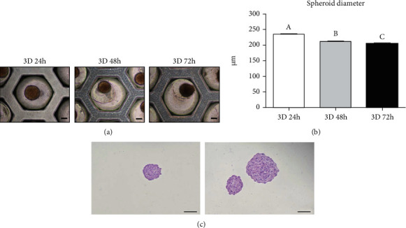
Formation of spheroids using DPSCs. (a) The aggregation of DPSCs into spheroids in 3D culture conditions (scale bar = 100 μm). (b) The DPSC spheroids became compact, and the diameter decreased with the time course of the 3D culture. Different superscripts (a to c) represent a significant (p < 0.05) difference. (c) H&E staining of the spheroid section from the 24-hour cultured group (scale bar = 200 μm).
